What Is The Retina?
The retina is a piece of tissue at the back of the eye. It is light-sensitive, transferring rays of light that pass through the front of the eye to the optic nerve. This is the pathway to the brain that results in vision. The retina is multi-layered. The outermost piece of tissue is made up of rods and cones that act as photoreceptors. Rods and cones transform light into electrical signals. At the center of the retina is the macula, which is responsible for central and detailed vision. As integral outgrowths of the developing brain, the optic nerve, and the retina are considered part of the central nervous system.
What Causes Retinal Diseases?
Retinal diseases can affect any part of your retina. The most common conditions that would qualify as a disease would be diabetic retinopathy, macular degeneration, and retinal detachment. Other conditions, such as a retinal tear or a macular hole can be the results of problems that lead to conditions like retinal detachment.
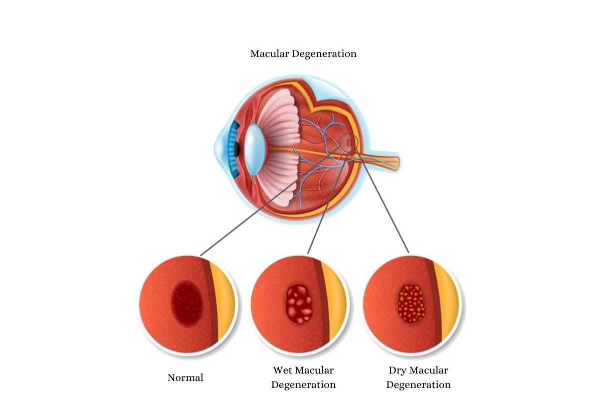
Age-related macular degeneration involves the breakdown of a small portion of the retina called the macula. The causes of macular degeneration are still somewhat a mystery, although it appears to be a combination of heredity and environmental factors, such as smoking, obesity, and diet.
Diabetic retinopathy involves the deterioration of blood vessels in the back of the eye that then leak fluid into and under the retina. This disease is caused by high blood sugar due to diabetes.
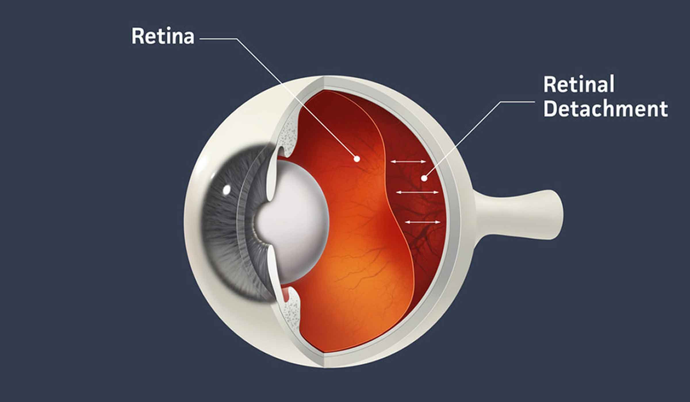
Retinal detachment happens when the retina pulls away from the back of the eye. This typically starts with a tear in the retina that allows fluid behind the retina, causing the retina to lift away from the underlying tissue layers. This is caused when the vitreous, the clear, gel-like substance in the center of the eye, shrinks, and tugs on the retina. This change in the vitreous happens with normal aging.
What Causes Flashes And Floaters?
Floaters look like small specks, dots, circles, cobwebs, or worm-like lines that appear out in front of your vision. They seem to be floating, hence the name. Actually, floaters are shadows being cast on your retina by the incoming light. The light is hitting tiny clumps of gel or cells inside the vitreous that fills our eyes and this casts a shadow onto the retina at the back of the eye. Floaters occur more as we age and the vitreous starts to thicken or shrink.
Flashes are also more common due to aging. They can look like a flashing light or a lightning streak in your field of vision. Flashes happen when the vitreous rubs or pulls on the retina, which is usually due to the shrinkage of the vitreous with age.
Neither floaters nor flashers are typically serious, but if you suddenly notice a change in the number of floaters or flashes, this can be a sign of retinal detachment and merits immediate attention from the team at North Suburban Eye Associates.
What Does The Retina Do?
The retina covers approximately 65 percent of the back of the eye. It is connected to the optic nerve, a part of the brain that receives electrical signals from the retina and transfers them to the brain. The retina is the part of the eye that receives light and transforms it into the signals necessary for vision.
What Are The Retina Diseases Treated At Georgina Eye Clinic?
We provide comprehensive care to patients experiencing retinal conditions. Two of the most commonly diagnosed problems include macular degeneration and detached retina.
What Can I Do To Prevent Retinal Diseases?
While there aren’t any specifics that can help you prevent macular degeneration or the changes that occur in the vitreous of your eyes that can lead to detachment, you can prevent diabetic retinopathy by avoiding the development of type 2 diabetes. Unlike type 1 diabetes, which is a congenital condition, type 2 diabetes can be prevented by maintaining your weight around your ideal body mass index, staying physically active, keeping your blood sugar levels down, and eating a more nutritionally sound diet.
What Are the Symptoms Of A Retinal Eye Disease?
Although the conditions affecting the retina vary, there is some commonality with the signs and symptoms. They may include:
- Suddenly seeing more floating specks or cobwebs (floaters)
- Blurred or distorted vision, where straight lines appear wavy
- Defects in peripheral vision
- Lost vision
Macular Degeneration
Often referred to as an age-related condition, macular degeneration (AMD) is the leading cause of vision loss and, at present, is incurable. Proper treatment is a critical aspect of preserving eyesight.
Macular degeneration is the deterioration of the center of the retina, the macula. The macula focuses on central vision and influences our ability to recognize faces, read, drive, and see fine details in objects. There are two types of macular degeneration that may develop: dry and wet types.

Dry macular degeneration is atrophic, meaning that the macula is suffering atrophy. The majority of cases of AMD are dry. The deterioration of the macula seems to be caused by the growth of small yellowish or white deposits called drusen beneath the macula.
Wet macular degeneration occurs in only 10 to 15% of cases. However, this type of macular degeneration accounts for most cases of severe vision loss. This form of macular degeneration is referred to as wet because it involves the growth of abnormal blood vessels that leak fluid and blood into the back of the eye. The accumulation of fluid can cause the retina to lift and separate from its base, resulting in vision loss.
How Soon Would I Need To See A Doctor For Macular Degeneration?
Macular degeneration can be spotted during your regular eye exams. We look for the appearance of drusen, yellow deposits on the retina. These indicate the development of early-stage dry macular degeneration. That reinforces the importance of maintaining regular eye exams once you pass the age of 40. From the ages of 40-54, you should have your eyes examined every two to four years. From 55 to 60, every one to three years, and every year after age 60.
If you haven’t been having regular exams, the first signs of increasing macular degeneration would be changes in your central vision and an impaired ability to see colors and fine detail. These issues would merit an immediate eye examination.
Retinal Detachment
Retinal detachment is a serious problem that can result in vision loss as the retina pulls away from the tissue around it, depriving it of nutrients and oxygen. This is an emergency that requires prompt medical attention.
When Is Surgery Necessary To Treat A Retinal Eye Disease?
The goal of the various treatments addressing different retinal diseases is to stop or slow the progression of the disease and to preserve, improve, or restore vision. With an inherited degenerative disease such as retinitis pigmentosa, the progression of the disease is quite slow, and there no cure or surgery that can stop the progression.
In other diseases, such as macular degeneration, management is key to protect the patient from losing more vision. Surgery for age-related macular degeneration could involve laser therapy to destroy abnormal blood vessels. Injections of medications, although not technically surgery, also come into play to stop the growth of new abnormal blood vessels.
In cases of retinal detachment, retinal tears, or macular holes, these conditions all require immediate surgery to keep the patient from losing vision.
What Are the Retina Treatments Offered At Georgina Eye Clinic?
Treatments for macular degeneration include:
- Anti-angiogenic medication. This is injected into the eye after the eye has been numbed with anesthetic drops. The medication prevents the growth of new abnormal blood vessels and also stops the leakage of blood and fluid from existing vessels.
- Laser therapy. In some cases, doctors may recommend treatment with a high-energy laser to destroy the abnormal blood vessels growing in the retina.

Treatments for retinal detachment include:
- Pneumatic retinopexy. This procedure stops the flow of fluid into the back of the retina by inserting an air or gas bubble into the vitreous cavity (the middle of the eye). The bubble presses the retina into place against the back wall of the eye, allowing fluid to absorb naturally over time. The bubble also absorbs on its own.
- Scleral buckling. This procedure relieves the pull on the retina by suturing a piece of silicone material to the sclera, the white of the eye. Buckling pushes the sclera toward the retina, creating appropriate pressure to seal a tear and prevent full detachment.
- Vitrectomy. The vitrectomy procedure drains the gel-like fluid that fills the center of the eye and replaces it with silicone oil, gas, or air. This replacement restores adequate pressure to secure the retina in place
Schedule A Consultation
Retinal problems can be managed with early intervention. To protect your long-term vision, contact Georgina Eye Clinic by calling 0755 347258 to schedule a consultation with one of our specialists
